


Breaking News
 DON MESS WITH ME Trump says Iran will 'be blown off face of earth' if they attack US warship
 Digital ID: Treasury Secretary Scott Bessent Reveals Trump Plans To Sign Executive Order To Hand...
 Pardon or not, there's one very dire reason why Fauci must be charged quickly…
 The Spanish-US Spat Could Lead To NATO's Unraveling
Top Tech News
 Robot Dives 1.5 Miles, Maps French Shipwreck With 86,000 Images And Recovers Artifacts
 Brain-inspired chip could reduce AI energy use by 70%
 CANCER HAS BEEN CURED
 "This is the first synthetic species," microbiologist J. Craig Venter told 60 Minutes'
 Humanoid robots are hitting the factories at an increasing pace
 Microsoft's $400 Billion Mistake Is Now a $200 Phone With Zero Tracking
 Turn Sand to Stone With Vinegar. Stronger Than Steel. Hidden Since 1627
 This is a bioprinter printing with living human cells in real time
 The remarkable initiative is called The Uncensored Library,...
 Researcher wins 1 bitcoin bounty for 'largest quantum attack' on underlying tech


3D color X-ray machine heads for trials
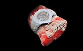
But this new scanner adds color and a third dimension, creating high resolution, cutaway 3D models that can diagnose bone fractures and monitor healing. New Zealand-based Mars Bioimaging (MBI) has now conducted a feasibility study of the machine, with a larger international trial set to begin soon.
In a traditional CT scan, X-rays are beamed through the target area of the body, and the radiation is absorbed more readily by denser tissues like bone, while passing more easily through softer tissues. The end result is that high contrast black-and-white image we know so well.
But the new technology collects more nuanced data about how the X-rays are absorbed by different tissues. It's built around a chip called the Medipix3, which tracks every photon that hits every pixel on the sensor, and processes their interactions with various atoms in the body. By doing so, it can determine the density and composition of those tissues more accurately.