


Breaking News
 Tina Peters, granted clemency by Colorado's Dem governor
 Tulsi Gabbard at center of explosive CIA claim as JFK and MKUltra files 'vanish from her office*
 Bessent Says US, China To Launch AI Safety Talks After Trump-Xi Meeting In Beijing
 Cuba Depletes Fuel As Blackouts Worsen, Putting Havana's Communists Under Pressure...
Top Tech News
 US To Develop Small Modular Nuclear Reactors For Commercial Shipping
 New York Mandates Kill Switch and Surveillance Software in Your 3D Printer ...
 Cameco Sees As Many As 20 AP1000 Nuclear Reactors On The Horizon
 His grandparents had heart disease.
At 11, Laurent Simons decided he wanted to fight aging.
His grandparents had heart disease.
At 11, Laurent Simons decided he wanted to fight aging.
 Mayo Clinic's AI Can Detect Pancreatic Cancer up to 3 Years Before Diagnosis–When Treatment...
A multi-terrain robot from China is going viral, not because of raw speed or power...
 The World's Biggest Fusion Reactor Just Hit A Milestone
 Wow. Researchers just built an AI that can control your body...
 Google Chrome silently installs a 4 GB AI model on your device without consent
 The $5 Battery That Never Dies - Edison Buried This 100 Years Ago


Magnetic resonance imaging of melanoma exosomes
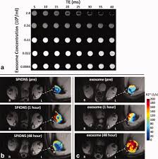
The ability to modify and track exosomes in vivo is essential to understanding exosome pathogenesis, and for utilizing exosomes as effective diagnostic and therapeutic nanocarriers to treat diseases.
Researchers from the Washington University School of Medicine recently reported a new electroporation method that allow exosomes to be loaded with superparamagnetic iron oxide nanoparticles for magnetic resonance tracking. Building on this approach, they now demonstrate for the first time using a C57BL/6 mouse model that melanoma exosomes can be imaged in vitro, and within lymph nodes in vivo with the use of standard MRI approaches.