










Breaking News
 Is Big Pharma Selling Postpartum Depression to New Moms?
 Make Men Men Again: Low T Is Now a National Defense Problem
 Druski: They're Lying To You About Overnight Success!
 $6.27 Diesel Just Hit Harvest. The Grocery Aisle Hasn't Felt It Yet.
Top Tech News
 Tesla Model 3 killer charges from 10 to 97% in just 9 minutes
 World-first unpowered DNA computer sets speed record
 I Power 5 Buildings Off-Grid. Here's How
 The US government is pushing hard to get a working nuclear fission reactor into space by late 2028
 SpaceX Starmind AI in Space Radiator
 China Will Dominate Global Nuclear Energy Through 2035, Analyst Says
 These Absolutely Wild-Looking EVs Are Saudi Arabia's First Homegrown Cars
 BEYOND THE MOON: NASA plans a nuclear-powered fleet to push DEEPER into space
 Big Oil Backs Mazama's $135 Million Bet On Superhot Geothermal
 Quantum compass takes flight on Northrop's Lumberjack drone


Tiny 3D-printed medical camera could be deployed from inside a syringe
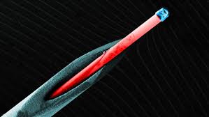
Getting inside the human body to have a look around is always going to be invasive, but that doesn't mean more can't be done to make things a little more comfortable. With this goal in mind, German researchers have developed a complex lens system no bigger than a grain of salt that fits inside a syringe. The imaging tool could make for not just more productive medical imaging, but tiny cameras for everything from drones to slimmer smartphones.


Scientists from the University of Stuttgart built their three-lens camera using a new 3D printing technique. They say their new approach offers sub-micrometer accuracy that makes it possible to 3D print optical lens systems with two or more lenses for the first time. Their resulting multi-lens system opens up the possibility of correcting for aberration (where a lens cannot bring all wavelengths of color to the same focal plane), which could enable higher image quality from smaller devices.
Here's how they did it. Using a femtosecond laser, where the pulse durations were shorter than 100 femtoseconds (a femtosecond is one quadrillionth of a second), they blasted a light-sensitive material resting on a glass substrate. Two photons are absorbed by the material, which exposes it and crosslinks polymers within. Unexposed material is then washed away with a solvent, leaving behind the hardened, crosslinked polymer used to form the optical element.
The team used this approach to print imaging components for optical microscopes with a diameter and height of 125 micrometers, and then attached them to the end of a 5.6-ft (1.7-m) optical fiber the width of two human hairs. The camera on the end of this small endoscope is capable of focusing on images from a distance of 3 mm (0.12 in). The team says the entire imaging system fits comfortably inside a standard syringe needle, which raises the possibility of delivering it to directly to organs, and even the brain.