


Breaking News
 Palantir Manifesto Shows The Clear Convergence Of Technofascism With Technocracy
 Washington's Democrat ex-governor says she's disgusted at millionaires' tax...
 The Odyssey Backlash Goes NUCLEAR - WTF Nolan?
 He Got Banned From Selling Skateboards | Joe Rogan
Top Tech News
 US To Develop Small Modular Nuclear Reactors For Commercial Shipping
 New York Mandates Kill Switch and Surveillance Software in Your 3D Printer ...
 Cameco Sees As Many As 20 AP1000 Nuclear Reactors On The Horizon
 His grandparents had heart disease.
At 11, Laurent Simons decided he wanted to fight aging.
His grandparents had heart disease.
At 11, Laurent Simons decided he wanted to fight aging.
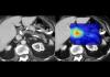 Mayo Clinic's AI Can Detect Pancreatic Cancer up to 3 Years Before Diagnosis–When Treatment...
A multi-terrain robot from China is going viral, not because of raw speed or power...
 The World's Biggest Fusion Reactor Just Hit A Milestone
 Wow. Researchers just built an AI that can control your body...
 Google Chrome silently installs a 4 GB AI model on your device without consent
 The $5 Battery That Never Dies - Edison Buried This 100 Years Ago


Reprogramming retina cells found to reverse blindness in mice
We owe our vision to an array of photoreceptor cells on our retinas, which respond to light and send the signals to the brain to interpret what we're seeing. But being neurons these cells won't regenerate on their own, so if they're damaged, that's it. At least, that's how it works in mammals – scientists have found that other animals like the zebrafish can convert structural cells called Müller glia into new, functioning photoreceptors to restore their vision. The new study has now shown how this could be done in mammals.
"This is the first report of scientists reprogramming Müller glia to become functional rod photoreceptors in the mammalian retina," says Thomas N. Greenwell, NEI program director for retinal neuroscience. "Rods allow us to see in low light, but they may also help preserve cone photoreceptors, which are important for color vision and high visual acuity. Cones tend to die in later-stage eye diseases. If rods can be regenerated from inside the eye, this might be a strategy for treating diseases of the eye that affect photoreceptors."
![]()
–– ADVERTISEMENT ––

The team investigated whether this kind of repair mechanism could be carried over to mammals, ideally without having to injure the retinas of test mice. Eventually they developed a two-phase process that managed to do just that. In the first phase, the researchers injected the eyes of healthy mice with a gene that would turn on a protein called beta-catenin. This triggers the Müller glia to start dividing. After a few weeks, phase two involved injecting factors into the eyes that direct those newly-divided cells to develop into rods.
When the team examined the cells using microscopy, they found that structurally the rods grown out of Müller glia looked exactly the same as the natural ones. On top of that, they also developed the network of synapses that allowed them to communicate with other neurons.