


Breaking News
 The Real Cost of Supporting the Zionist Secular State of Israel
 Treasury Yield 30 Years (^TYX)
 Apoplectic Netanyahu rages at Trump in private as 'disastrous' Iran deal leaves him longing.
 The American Consumer Is Piss Broke
Top Tech News
 Elon and SpaceX Have Made AI Training 10 Times Faster
 Oklo COO Says Nuclear Waste Could Power America For 150 Years
 SpaceX Announces LARGEST Starship Mission Ever! They've never done this before!
 Cars Are Fast Becoming Dystopian Prison Pods...
 Our Emergency Water Plan Wasn't Good Enough - So We Built This
 Sodium Ion Batteries Can Reach 100 Gigawatt Per Hour Per Year Scale in 2027
 Juiced Bikes proves capable electric motorcycles don't have to cost a lot
 Headlight projectors turn your car into a drive-in theater
 US To Develop Small Modular Nuclear Reactors For Commercial Shipping
 New York Mandates Kill Switch and Surveillance Software in Your 3D Printer ...


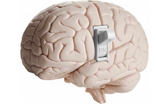
'Sugar switch' in the brain offers new path to treating depression
Scientists from South Korea's Institute for Basic Science (IBS) found that prolonged stress changes how proteins in the medial prefrontal cortex (mPFC) are "decorated" with sialic acid – a sugar molecule that helps shape the surface properties of neurons. These sugar chains, called glycans, are attached after proteins are made, forming the process known as glycosylation. Glycosylation has been studied in terms of how it impacts cancer development and, more recently, neurodegeneration.
One type of glycosylation is O-glycosylation, where sugars attach to oxygen atoms on certain amino acids in a protein. This molecular "sugar coating" helps regulate how neurons connect and signal to one another. Until recently, it was largely overlooked in mental-health research, but scientists are now finding that stress can disturb these sugar patterns, potentially rewiring "normal" communication between brain cells.
In this study, the researchers identified that a single enzyme, St3gal1, performs the final "sugar-capping" step in the process, and this small but integral stage influences how long proteins last and how they interact at synapses – and how, if this falters, it appears to influence depression-like behaviors as a result.
Researchers used high-performance mass spectrometry to map O-glycosylation patterns across nine brain regions in healthy mice. Each area had a distinct sugar signature, reflecting its unique cellular blueprint. When the team compared these with those from chronically stressed mice, they found a significant difference in the prefrontal cortex, a region linked to mood regulation. Here, stress led to a noticeable reduction in that final O-glycosylation sugar-capping step, and a corresponding drop in St3gal1 expression.
Knocking out St3gal1 in healthy mice caused depressive symptoms, including loss of motivation and heightened anxiety. Increasing St3gal1 in stressed mice had the opposite effect, easing those behaviors. This showed that the enzyme has a key role in how stress triggers depression-like changes in the brain. The team also found that St3gal1 helps maintain sugar tags on neurexin-2, a protein that supports communication between neurons. In stressed mice, those tags vanished – along with normal neural signaling – but restoring St3gal1 brought them back.
"This study demonstrates that abnormal glycosylation in the brain is directly connected to the onset of depression," said research fellow Boyoung Lee. "It provides an important foothold for identifying new diagnostic markers and therapeutic targets beyond neurotransmitters."
While the findings were only demonstrated in the brains of male mice – and, of course, the neural networks in humans are far more complex – it provides a new angle for research into depression and its treatment. Many current antidepressants act on serotonin – boosting its levels or altering its signaling – but there's growing evidence suggesting that it's not simply a case of "too little serotonin."