


Breaking News
 The Real Cost of Supporting the Zionist Secular State of Israel
 Treasury Yield 30 Years (^TYX)
 Apoplectic Netanyahu rages at Trump in private as 'disastrous' Iran deal leaves him longing.
 The American Consumer Is Piss Broke
Top Tech News
 Elon and SpaceX Have Made AI Training 10 Times Faster
 Oklo COO Says Nuclear Waste Could Power America For 150 Years
 SpaceX Announces LARGEST Starship Mission Ever! They've never done this before!
 Cars Are Fast Becoming Dystopian Prison Pods...
 Our Emergency Water Plan Wasn't Good Enough - So We Built This
 Sodium Ion Batteries Can Reach 100 Gigawatt Per Hour Per Year Scale in 2027
 Juiced Bikes proves capable electric motorcycles don't have to cost a lot
 Headlight projectors turn your car into a drive-in theater
 US To Develop Small Modular Nuclear Reactors For Commercial Shipping
 New York Mandates Kill Switch and Surveillance Software in Your 3D Printer ...


The Immense Complexity of a Brain is Mapped in 3D for the First Time:
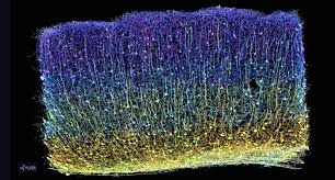
During the last seven years, a global team of more than 150 scientists collaborated on the most complicated neuroscience experiment ever attempted—and they've released their findings this week.
From a tiny sample of tissue no larger than a grain of sand, the MICrONS Project completed the first step toward the goal once thought unattainable: building a functional wiring diagram of a portion of the brain.
Now, they've published their findings in Nature with a collection of ten studies. The 3D wiring diagram and its data are massive—1.6 petabytes in size (equivalent to 22 years of non-stop HD video). They offer a never-before-seen insight into brain function and organization of the visual system.
The research started at Baylor College of Medicine where scientists used specialized microscopes to record the brain activity from a one cubic millimeter portion of a mouse's visual cortex while the animal watched various movies and YouTube clips.
Afterwards, Allen Institute researchers took that same cubic millimeter of the brain and shaved it into more than 25,000 layers, each 1/400th the width of a human hair, and used an array of electron microscopes to take high-resolution pictures of each slice.
By the end, the MICrONS Project—Machine Intelligence from Cortical Networks—built the most detailed wiring diagram of a mammalian brain to date—and it's freely available online.
"A watershed moment for neuroscience, comparable to the Human Genome Project" is the description from David Markowitz, Ph.D., who coordinated this work after leaving the IARPA, the US Intelligence Advanced Research Projects Activity, which partially funded it.
Another team at Princeton University used artificial intelligence and machine learning to reconstruct the cells and connections into a 3D volume. Combined with the recordings of brain activity, it contains 523 million synapses (the connection points between 200,000 cells) and a length of four kilometers of axons (the branches that reach out to other cells).
"Inside that tiny speck is an entire architecture like an exquisite forest," said Clay Reid, Ph.D., senior investigator and one of the early founders of electron microscopy connectomics who brought this area of science to the Allen Institute 13 years ago.
"It has all sorts of rules of connections that we knew from various parts of neuroscience—and within the reconstruction itself, we can test the old theories and hope to find new things that no one has ever seen before."