


Breaking News
 Palantir Manifesto Shows The Clear Convergence Of Technofascism With Technocracy
 Washington's Democrat ex-governor says she's disgusted at millionaires' tax...
 The Odyssey Backlash Goes NUCLEAR - WTF Nolan?
 He Got Banned From Selling Skateboards | Joe Rogan
Top Tech News
 US To Develop Small Modular Nuclear Reactors For Commercial Shipping
 New York Mandates Kill Switch and Surveillance Software in Your 3D Printer ...
 Cameco Sees As Many As 20 AP1000 Nuclear Reactors On The Horizon
 His grandparents had heart disease.
At 11, Laurent Simons decided he wanted to fight aging.
His grandparents had heart disease.
At 11, Laurent Simons decided he wanted to fight aging.
 Mayo Clinic's AI Can Detect Pancreatic Cancer up to 3 Years Before Diagnosis–When Treatment...
A multi-terrain robot from China is going viral, not because of raw speed or power...
 The World's Biggest Fusion Reactor Just Hit A Milestone
 Wow. Researchers just built an AI that can control your body...
 Google Chrome silently installs a 4 GB AI model on your device without consent
 The $5 Battery That Never Dies - Edison Buried This 100 Years Ago


CERN chip enables first 3D color X-ray images of the human body
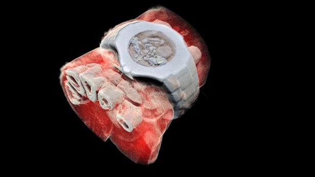
Medical X-ray scans have long been stuck in the black-and-white, silent-movie era. Sure, the contrast helps doctors spot breaks and fractures in bones, but more detail could help pinpoint other problems. Now, a company from New Zealand has developed a bioimaging scanner that can produce full color, three dimensional images of bones, lipids, and soft tissue, thanks to a sensor chip developed at CERN for use in the Large Hadron Collider.
Mars Bioimaging, the company behind the new scanner, describes the leap as similar to that of black-and-white to color photography. In traditional CT scans, X-rays are beamed through tissue and their intensity is measured on the other side. Since denser materials like bone attenuate (weaken the energy) of X-rays more than soft tissue does, their shape becomes clear as a flat, monochrome image.

But for the new technology, which Mars calls "Spectral CT," the sensor can measure the attenuation of specific wavelengths of the X-rays as they pass through different materials. After running the spectroscopic data through specific algorithms, a 3D color image is generated that clearly shows muscle, bone, water, fat, disease markers – and even a watch. The end results are unnerving, like someone's sculpted a detailed clay model of your insides.
At the heart of the Spectral CT scanner is a Medipix3 chip. This device, which detects and counts every individual particle that hits each pixel on the sensor, was originally developed at CERN to precisely track particles in the Large Hadron Collider.
A small version of the device has been tested to see how well it can diagnose bone and joint health, spot cancer, and pick up early markers for vascular diseases. So far, the results have been promising, the team says.
"In all of these studies, promising early results suggest that when spectral imaging is routinely used in clinics it will enable more accurate diagnosis and personalization of treatment," says Anthony Butler, one of the creators of the 3D scanner.